Camponotus ligniperda
The biggest Slovak ant
Techical note: This is only word to word translate.
Camponotus ligniperda popularly called ant fungi, decay is one of the largest European species. On the territory of Slovakia is our largest species. Workers can grow to the size of 15mm. Dogs are stained black with dark red chest. Occurs on sunny slopes and dry plains, where they build nests in wood or under stones. Live the sweet secretions from aphids and small insects.
The queen, after mating head seeks refuge in the gaps of wood. She then founded a colony. The eggs laid by the hatched larvae, cocoons and then create a newly hatched workers then provide for the queen. ligniperda Camponotus creates caste major and minor. The first workers are small stature. Their development takes aproximatly 50 days. Later start seeing major.
Camponotus ligniperda can be for its size simply observe and photograph. Next followed a collection of pictures that I've accumulated. I used the macro, light and electron microscopy. Images are higher resolution, so it may be somewhat hindered.
Macrophotography
I placed the camera formikáriu, so I can respond rapidly when something interesting happens. The problem resides in the large aperture (ie small aperture opening) is too little light, so I keep photos sharp. Sometimes, but are lucky enough and ant poses the right light. The photo below is not from nature, but behind glass formicary.
.
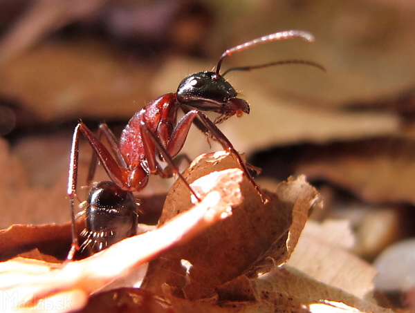
Workers in a ferocious attitude.
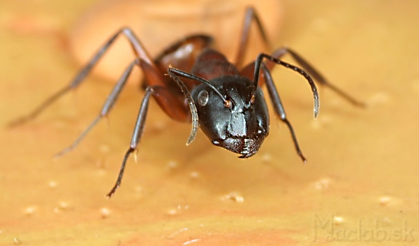
Detail of prepared worker.
Ctlick into the image to rotate ant of 360° angles
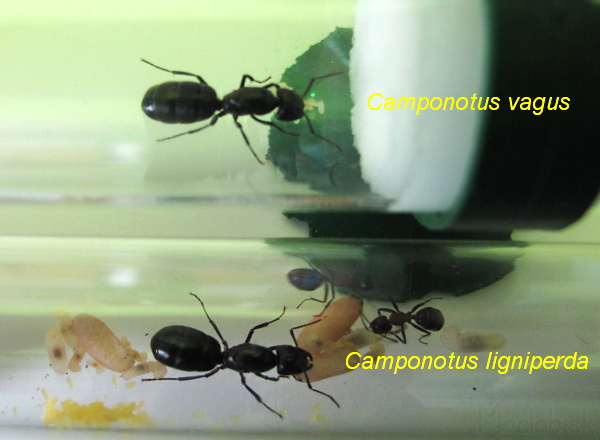
For comparison, two queens side by side.
Light microscopy
The light microscope has very little depth of field, but getting it colored pictures. The ant is so big and rugged, that can not be used or a method of software extension depth of field when more images combined into one.
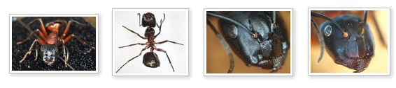
Photos by light microscopy. Click to enlarge.
Electron microscopy
SEM (by scanning electron microscopy) - scanning electron microscopy uses a beam of electrons instead of light. Depth of field is very large, but the resulting image is only in shades of gray. The SEM can be observed only conductive materials, otherwise would have naprášiť carbon.
Observation of ant I used the method of GBL (Gentle low beam), which allows you to watch and nonconductive samples. Accelerating voltage up to 1kV and is also on the sample holder is introduced negative bias. Bias repels electrons that would otherwise accumulate on the surface of non-conductive samples.
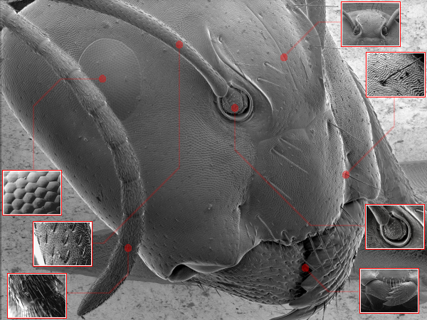
Click on the rectangle to zoom.
Press F11 to toggle full-screen viewing.
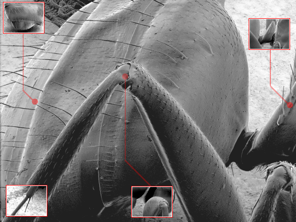
Click on the rectangle to zoom.

Download picture in high quality. Click to select the resolution.
In the future I plan to :
- capture 3D stereographic image
- capture many obrzázkov rotating ant, so that it could re-play from every angle
- done using ion beam cross section and look at the ant from the inside
Where to go?
Production of formicary from Ytong (not for only Camponotus ligniperda)
![]()
Photographs of plants were created at HRSEM JEOL 7600F + WDS + EDS + EBSD
Centre for Development and application of advanced diagnostic methods in the processing of metallic and nonmetallic materials
ITMS 26220120014